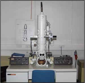
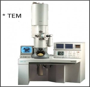
Electron microscope is a instrument used to study the fine details of cell structure by which magnification is done by emitting a electron beam at high voltage through electromagnatic lenses in a vaccum chamber.
There are two types of electron microscopes; Transmission electron microscope (TEM) and Scanning electron microscope. In Transmission electron microscope, the electron beam passes through the specimen and projects an image onto a fluorescent screen (Zinc sulphide). It magnifies the image up to 500,000 times than its normal size. Its resolution is 0.14nm. The Transmission electron microscope is used to study the two dimentional features of intrastructure (ultra structure) of the specimen. Ultra thin sections (60nm) are examined with Transmission electron microscope.
In Scanning electron microscope, Incident electron beam onto the specimen will be reflected and form an image on TV screen. It magnifies the image 200,000 times. Its resolution power is 5nm. It scans the surface of the specimen and gives detailed surface structure. Biological and non biological samples are examined using scanning electron microscope.
The ideal specimen for electron microscopy is biopsies. Living tissues can not be examined with Electron microscope. Therefore the tissue should be preserved as closely as living state. 2{58cd716989699d561e07eed7f371f717b4e2b8d0a4dba95f1ca9150bd3161838} Gluteraldehyde is known as good fixative for preserving electron microscopic specimens. The biopsy collected for electron microscope should be placed at once in 2{58cd716989699d561e07eed7f371f717b4e2b8d0a4dba95f1ca9150bd3161838} gluteraldehyde containing bottle and sent to the laboratory in ice as soon as possible. If delay is unavoidable, the specimen should be stored at 4oC. The 5×5 mm size biopsy is cut into 4-5 pieces and washed in 0.1{58cd716989699d561e07eed7f371f717b4e2b8d0a4dba95f1ca9150bd3161838} PBS for 5-6 times. each wash should take at least 5 minutes.Then the specimen should be treated with 1{58cd716989699d561e07eed7f371f717b4e2b8d0a4dba95f1ca9150bd3161838} osmium tetra oxide for 10 minutes.Osmium tetra oxide is highly carcinogenic. Therefore this procedure should be done under a fume hood.
After treatment , the specimen is washed in PBS for 45 minutes to 1 hour. Next, the specimen should be sent through the alcohol series (50{58cd716989699d561e07eed7f371f717b4e2b8d0a4dba95f1ca9150bd3161838}, 60{58cd716989699d561e07eed7f371f717b4e2b8d0a4dba95f1ca9150bd3161838}, 70{58cd716989699d561e07eed7f371f717b4e2b8d0a4dba95f1ca9150bd3161838}, 80{58cd716989699d561e07eed7f371f717b4e2b8d0a4dba95f1ca9150bd3161838}, 90{58cd716989699d561e07eed7f371f717b4e2b8d0a4dba95f1ca9150bd3161838},and Two absolute alcohols). In each alcohol, the specimen should be kept for alcohol. Then place the specimen in gran alcohol which contains CUSO4. Then the specimen is washed in PBS for 10 minutes. The specimen is treated with propylene oxide twice. each treatment should be for 15 minutes.
100{58cd716989699d561e07eed7f371f717b4e2b8d0a4dba95f1ca9150bd3161838} Resin is gradually poured in to the embedding mould without forming air bubbles and the processed biopsy is placed on the top of resin. At least for blockes should be prepared from one specimen. Then the mould is placed in the 35oCoven overnight. The mould should be placed at 45oC for 4-7 hours and then it should be kept at 60oC overnight. Then the resin embedded tissue is removed from mould and semi thick sections are cut using electron microtomy for find the good block. For that a drop of distilled water is placed on the slide and then semi sectioned specimen is placed on the drop using forcep. The resin is allowed to evaporate and the smear is stained with 1{58cd716989699d561e07eed7f371f717b4e2b8d0a4dba95f1ca9150bd3161838} Toludine Blue for 2 minutes. Then the slide should be washed and allowed to dry. The good block is choosen by seeing the sections of each block under the microscope.
The good block is placed in the microscope and adjusted the sectioning size to 60nm. The sections are cut by diamond knife and they are floated in the boat shaped structure which is located in front of knife. The sections are taken into nickel or copper mesh. Then the section is stained heavy metal ion containing SATO solution which contains Urenial acetate, Lead citrate, Lead nitrate and Lead acetate. These stained sections are examine through the electron microscopy.

dear
i saw ur note it is very nice.i am following a Msc in clinical biochemistry.try to do the same thing again
Thank you.
I’ll try to post related articles frequently. Keep on reading.